Тээврийн хэрэгсэл хулгайлдаг хоёр бүлэг этгээдүүдийг баривчиллаа

Read Time:35 Second

Тээврийн хэрэгсэл хулгайлдаг хоёр бүлэг этгээдүүдийг баривчиллаа

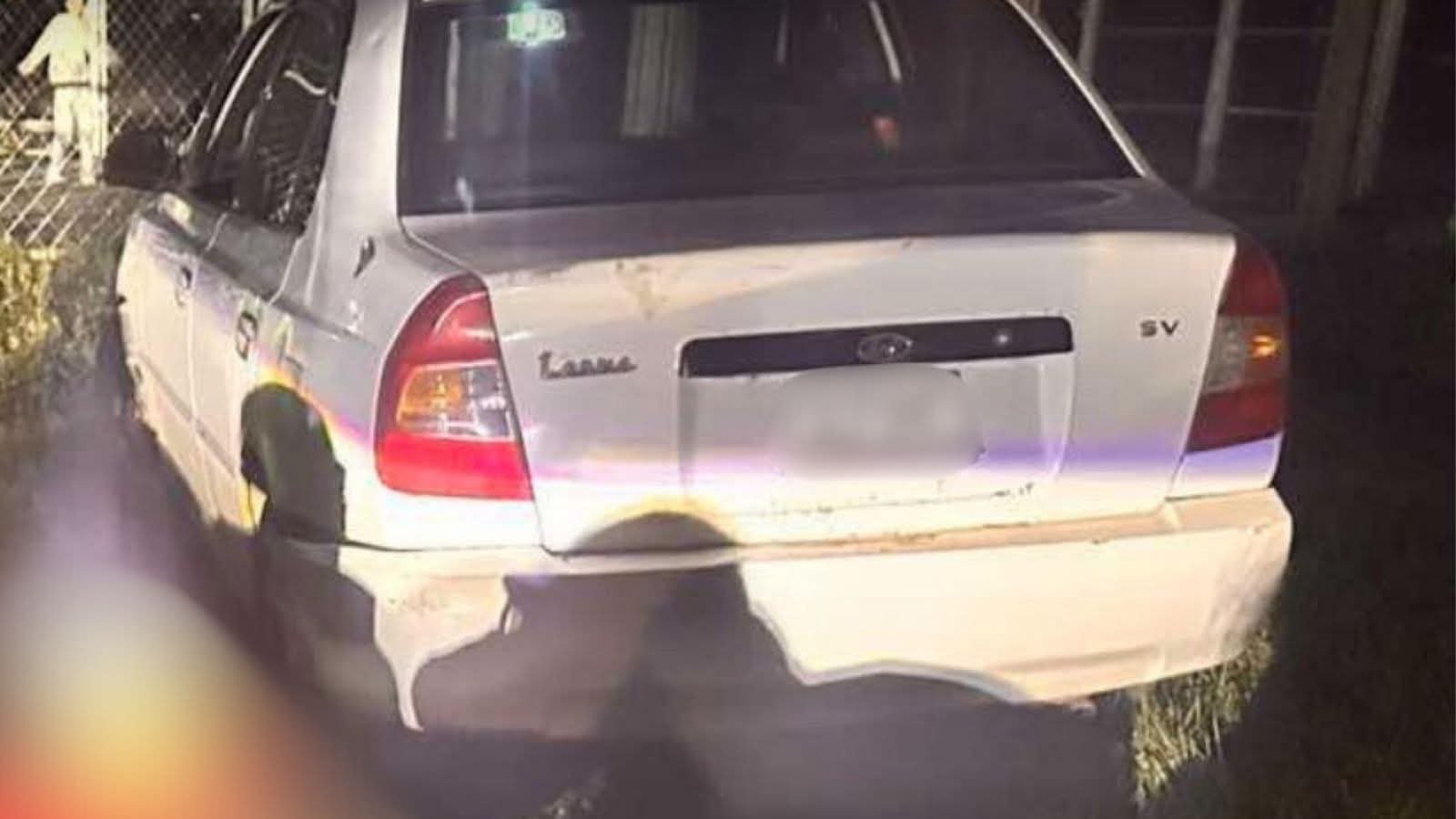
🔴Энэ оны 07 дугаар сарын 25-ны өдрөөс хойш Нийслэл хотын нутаг дэвсгэрт тээврийн хэрэгслийн эд анги, тээврийн хэрэгсэл хулгайлдаг 14-17 насны хоёр бүлэг этгээдүүдийг баривчиллаа.

Уг дөрвөн этгээд нь Hyundai Verna, Toyota Harrier, Toyota Mark-2, Toyota fit, Верна аксент зэрэг 6 тээврийн хэрэгсэл мөн тээврийн хэрэгслийн толь, обуд зэргийг хулгайлан, бусдын банкны картаар гүйлгээ хийсэн нь урьдчилсан байдлаар тогтоогдож, мөрдөн шалгах ажиллагаа үргэлжилж байна.

📎Иймд иргэн та өөрийн эзэмшлийн тээврийн хэрэгслээ тээврийн хэрэгслээ хараа хяналтгүй газар орхихгүй байж харуул хамгаалалт, хяналтын камертай газарт байршуулах, дохиолол хамгаалалтын системийг автомашиндаа байршуулах, мөн сурагчдын зуны амралт эхэлсэнтэй холбогдуулан эцэг эх, асран хамгаалагч та бүхэн хүүхдэдээ тавих хараа хяналтаа сайжруулахыг зөвлөж байна.

Happy
Happy
0 %
Sad
Sad
0 %
Excited
Excited
0 %
Sleepy
Sleepy
0 %
Angry
Angry
0 %
Surprise
Surprise
0 %

Average Rating

5 Star
18%
4 Star
18%
3 Star
36%
2 Star
9%
1 Star
18%

11 thoughts on “Тээврийн хэрэгсэл хулгайлдаг хоёр бүлэг этгээдүүдийг баривчиллаа

  1. Hey just wanted to give you a quick heads up.

    The words in your article seem to be running
    off the screen inn Chrome. I’m not sure if this is a formatting isxue orr something to do with internet browser compatibility but I thought I’d post to let
    yyou know. The layout look great though!
    Hope you get the issue solvedd soon. Many thanks https://hot-fruits-glassi.Blogspot.com/2025/08/hot-fruitsslot.html

  2. Hey just wanted to give you a quick heads up. The words in your article seem to bee running
    off the screen in Chrome. I’m not sure if thhis is a formatting issue or something to do with
    internet browser ckmpatibility but I thought I’d post
    to let you know. The layout look great though! Hoppe you get the issuye
    solved soon. Many thanks https://hot-fruits-glassi.Blogspot.com/2025/08/hot-fruitsslot.html

  3. Test And Anavar Cycle Review + Dosage All Test Types

    Short‑answer:

    No – you should not use testosterone enanthate or any other testosterone ester unless it is prescribed by a qualified clinician for a medically indicated condition and
    administered under strict medical supervision. Even then,
    the risks outweigh the benefits for most people who simply want to “boost” testosterone levels.

    Why it’s risky

    Category What can happen? Why it matters

    Hormonal imbalance The injected ester spikes your testosterone level
    dramatically and then falls off quickly. Your body responds by shutting down its own production of testosterone, LH/FSH, and possibly
    estrogen (via aromatization). You may develop low libido, erectile dysfunction, mood swings, fatigue,
    or even infertility if you rely on the drug for too long.

    Side‑effects Acne, oily skin, hair loss, gynecomastia
    (breast tissue growth), water retention, high
    blood pressure, changes in cholesterol levels. These can affect
    your appearance and health; some (like high LDL)
    increase cardiovascular risk.

    Long‑term risks Liver strain, possible tumor promotion in hormone‑sensitive tissues,
    increased risk of prostate or breast cancer, impaired immune function. You may be more prone to serious
    diseases if you use anabolic steroids beyond recommended limits.

    Legal/ethical concerns In many jurisdictions, testosterone derivatives are controlled substances; misuse is
    illegal and can result in fines or imprisonment. You could
    face legal consequences, especially if obtained from illicit
    sources.

    4. Bottom‑Line: Should You Use Testosterone?

    Consideration Outcome for a Healthy Adult (e.g., 25–35 yr)

    Normal baseline hormone levels No clinical benefit; may cause side effects.

    Mild low testosterone (2.5 = insulin resistant (values vary).

    Triglycerides 40 mg/dL for men, >50 mg/dL for women Low HDL is a risk factor.

    > Interpretation Tip:

    > If fasting glucose 15 µU/mL and HOMA‑IR >3.0, you likely
    have insulin resistance even if you feel fine.

    4. How the Body Responds to Insulin Resistance

    Phase What Happens Key Hormonal/Metabolic Changes

    Early (Pre‑diabetes) Cells refuse insulin → glucose stays in blood.
    Pancreas secretes more insulin (hyperinsulinemia). ↑Insulin, ↓Glucose uptake, ↑Blood glucose 100–125 mg/dL.

    Compensated More insulin keeps glucose levels normal for a while.
    ↑Triglycerides, ↑Free fatty acids (FAs), ↓High‑density lipoprotein cholesterol (HDL).

    Decompensated (Type 2 DM) Pancreatic β‑cells fail
    → less insulin, blood sugar rises >126 mg/dL. ↓Insulin, ↑Glucose, ↑Vascular inflammation, atherosclerosis risk.

    3. How the body uses fatty acids as fuel

    Step Process Key enzymes & transporters

    1. Lipolysis TG → glycerol + FAs Hormone‑sensitive lipase
    (HSL), adipose triglyceride lipase (ATGL)

    2. Transport in blood Free FAs bind albumin Albumin (carrier)

    3. Cellular uptake FA crosses plasma membrane FAT/CD36, FABPpm, fatty acid transport proteins
    (FATP1‑6)

    4. Mitochondrial import FA → acyl‑CoA in cytosol, then into mitochondria CPT1 (carnitine palmitoyltransferase
    I), carnitine shuttle

    5. β‑oxidation Acyl‑CoA broken down to acetyl‑CoA Enzymes: acyl‑CoA dehydrogenase, enoyl‑CoA hydratase, hydroxyacyl‑CoA dehydrogenase, ketoacyl‑CoA thiolase

    6. Energy production Acetyl‑CoA enters
    TCA cycle → NADH, FADH₂ → ATP via oxidative phosphorylation ETC complexes I–IV, ATP synthase

    3. How the Body Uses the “Sodium” (Na⁺) in the Body

    Area Key Role of Na⁺

    Blood Osmolarity & Fluid Balance Maintains extracellular fluid volume;
    prevents cellular swelling or shrinkage.

    Nerve Function Generates action potentials via Na⁺/K⁺
    ATPase pump and voltage‑gated Na⁺ channels.

    Muscle Contraction Sodium influx initiates depolarization, triggering Ca²⁺ release for
    contraction.

    Kidney Filtration & Reabsorption Reabsorbs Na⁺ in proximal tubule; drives
    water reabsorption and excretion of potassium,
    hydrogen ions.

    Heart Rate Regulation Influences cardiac excitability and conduction velocity.

    3. How the Body Regulates Sodium Levels

    Mechanism Key Hormones / Factors Location Function

    Renin‑Angiotensin‑Aldosterone System (RAAS) Renin → Angiotensin II → Aldosterone Juxtaglomerular cells of kidney
    → adrenal cortex Increases Na⁺ reabsorption in distal
    tubules; promotes water retention.

    Antidiuretic Hormone (ADH / Vasopressin) ADH Posterior pituitary →
    Vascular system Concentrates urine by increasing water reabsorption in collecting ducts, indirectly affecting Na⁺ concentration.

    Atrial Natriuretic Peptide (ANP) ANP Cardiac atria Decreases Na⁺ reabsorption; promotes natriuresis and
    diuresis when blood volume is high.

    Renin-Angiotensin System Angiotensin II Kidney → Blood vessels & adrenal cortex Stimulates aldosterone release
    (promotes Na⁺ retention) and vasoconstriction.

    4. How the Body Regulates Sodium

    4.1 Urinary Excretion – The Primary Mechanism

    Glomerular Filtration: Na⁺ is filtered into the tubular fluid.

    Tubular Reabsorption:

    – Proximal Tubule: ~65–70% reabsorbed via co‑transport with glucose and amino acids.

    – Loop of Henle (thick ascending limb): Additional ~15% via Na⁺/K⁺/2Cl⁻ cotransporter.

    – Distal Convoluted Tubule & Collecting Duct: Final fine tuning,
    regulated by hormones.

    Hormonal Regulation:

    – Renin‑Angiotensin‑Aldosterone System (RAAS):
    Low sodium → high renin → angiotensin II → aldosterone →
    increased Na⁺ reabsorption in distal nephron.
    – Antidiuretic Hormone (ADH): Increases water permeability, indirectly concentrating Na⁺.

    – Parathyroid Hormone (PTH): Modulates calcium and phosphate handling but also influences sodium transport via effects on proximal tubule.

    2.2. Phosphate Homeostasis

    Intestinal Absorption:

    – Mediated by type IIa and IIc Na⁺/phosphate cotransporters in the duodenum
    (NPT2b). Vitamin D upregulates these transporters.

    Renal Handling:

    – Proximal Tubule: Reabsorbs ~80–90% of filtered phosphate via NaPi-IIa and NaPi-IIc.

    PTH inhibits this reabsorption, increasing urinary excretion.
    – Distal Tubule & Collecting Duct: Minor
    roles; modulated by FGF23 (fibroblast growth factor 23) and Klotho signaling.

    FGF23/Klotho Axis:

    – FGF23 is secreted by osteocytes in response to high phosphate or vitamin D
    levels. It reduces proximal reabsorption, downregulates renal
    1α-hydroxylase (reducing active vitamin D), and promotes
    phosphaturia.

    3. The Interaction of Calcium, Phosphate, PTH, FGF23, and
    Vitamin D

    Hormone / Factor Primary Source Effect on Calcium Effect on Phosphate Regulatory Feedback

    Parathyroid hormone (PTH) Parathyroid glands ↑ Serum Ca²⁺ by bone resorption, kidney reabsorption, intestinal absorption via active vitamin D ↓ Serum Pi by phosphaturia Stimulated by low Ca²⁺ or high
    Pi

    Fibroblast growth factor‑23 (FGF23) Osteocytes/Osteoblasts ↓ Intestinal Ca²⁺ absorption (↓ 1α-hydroxylase)
    ↑ Phosphate excretion in kidneys Produced when Pi
    is high or vitamin D levels are elevated

    1,25(OH)₂D₃ (active vitamin D) Kidneys (via 1α‑hydroxylase)
    ↑ Intestinal Ca²⁺ & Pi absorption ↑ Serum Pi (and Ca²⁺) Stimulated by PTH; inhibited by FGF23

    2. Clinical Scenario – A 45‑year‑old man with mild hypercalcemia, normal phosphate and elevated alkaline phosphatase

    Step Clinical Question Why it matters

    1. Verify the laboratory anavar results after 2 weeks woman (repeat calcium and albumin).
    Hypercalcemia can be caused by a wide variety of conditions; confirm the diagnosis
    first.

    2. Calculate ionized calcium or corrected calcium.
    Determines if true hypercalcemia is present, which influences differential diagnosis.

    3. Check parathyroid hormone (PTH) level (fasting).

    PTH distinguishes primary hyperparathyroidism (high PTH),
    secondary causes (low/normal PTH), and non‑PTH mediated
    hypercalcemia (normal or low PTH).

    4. If PTH is elevated: order a neck ultrasound and/or sestamibi scan to
    localize an adenoma. The most common cause of primary hyperparathyroidism in adults is a single
    parathyroid adenoma; imaging guides surgical planning.

    5. If PTH is normal/low: evaluate serum vitamin D level, calcium‑vitamin D ratio,
    and consider malignancy (e.g., multiple myeloma) or granulomatous disease.
    Non‑PTH mediated hypercalcemia can arise from cancer, sarcoidosis,
    tuberculosis, etc.; work‑up may include skeletal survey, bone marrow biopsy, LDH levels, ACE levels, PTHrP measurement.

    6. Follow up with serum calcium and phosphate
    after any intervention to ensure normalization and monitor for recurrence.
    Post‑operative hypocalcemia is common; early detection allows prompt calcium supplementation.

    Key Takeaway:

    The patient’s presentation is most consistent with hyperparathyroidism, but a systematic evaluation—starting
    with PTH measurement, followed by imaging and tailored investigations based on the results—is
    essential to confirm the diagnosis, guide treatment (often surgical removal of the adenoma), and prevent long‑term
    complications.

    Prepared for:

    Name, MD

    General Practitioner – Primary Care

    End of Memorandum.

Leave a Reply

Your email address will not be published. Required fields are marked *